Texas Nerve and Paralysis Institute


Before After
Before and After Mod Quad Surgery. Over a Period of 5 Years. Abduction.
2001-1-12006-8-23
*Click on a picture to visit the photogallery.
Close
Dr. Nath - Brachial Plexus injury expert specializing in erbs palsy and brachial plexus palsy treatment.
CHILDREN WITH OBSTETRIC BPI ADULT OBSTETRIC & TRAUMATIC BPI PATIENTS SEEN ELSEWHERE
VISITING INTERNATIONAL SCHOLAR PROGRAM
Texas Nerve and Paralysis Logo - TNPI The Next Generation of Healthcare Portal on the Internet. Dr. Nath'S Pics
Dr. Nath's calendar Dr. Nath's forum Dr. Nath's Weblog Dr. Nath's Location - Texas Nerve and Paralysis Institute. Contact Dr. Nath Search Dr. Nath Brachial Plexus Website
Click above to Contact LOCATION


2201 W. Holcombe Blvd.
Suite 225
Houston , TX 77030
[ Map ]
Toll-Free
(866) 675-2200

Telephone
(713) 592-9900

Fax
(713) 592-9921


Dr.Nath's eNewsletter
View Archives
Sign Up  


Brachial Plexus Forum

Share ideas, lifestyle,
techniques interesting
observations and any other
issues you wish to discuss


PHYSICIAN AND
  THERAPIST’S ONLINE
  REFERRAL SYSTEM

For therapists,doctors
and other health care
providers who wish to
reffer patients for
evaluation and/or treatment.


Brachial Plexus Art
  Gallery

A Gallery of images
showing the human side
of the injury, and
giving sense of hope
for the future.


The Brachial Plexus
  Store

Colorful, uniques items
that show your support
for research in Brachial
Plexus injury.100 percent
of proceeds go to
charitable organisations.


Nerve Surgery Illustrations & Animations

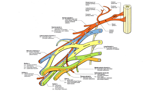
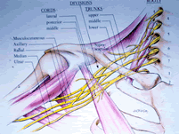
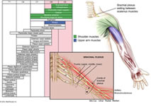
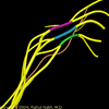
Brachial plexus anatomy is complex, but can be described in terms of basic function. The brachial plexus is derived from 5 "roots" or spinal nerves that exit the spinal cord in the neck. The nerves then pass through the axilla or armpit behind the collarbone (clavicle), and split into the final nerve branches that supply the muscles and skin of the shoulder, arm, elbow and hand.

The roots are named for the level of spinal cord that they exit. The upper roots (C5 and C6) exit from the cervical (meaning "neck") 5th and 6th vertebrae. The middle root (C7) exits above the 7th cervical vertebrae, and the lower roots exit from C8 (below the 7th cervical vertebra) and T1 (below the 1st thoracic vertebra). The upper roots supply upper structures (C5 to shoulder, C6 and C7 to elbow), and the lower roots supply the forearm and hand.

Brachial plexus anatomy


Injury to upper roots (the most common injury) is known as Erb’s palsy after the physician who described it. Erb’s palsy (paralysis) affects the shoulder and elbow, because the upper roots supply these structures. Injury to the lower roots is known as Klumpke’s paralysis, but is relatively uncommon, and very rarely exists by itself. Because the lower roots are injured, the hand is predominantly affected. If the lower roots are injured, the injury is generally so severe that all the roots of the plexus are involved, and the injury includes all parts of the arm. Upper root (Erb’s) injury is seen about 60% of the time, isolated Klumpke’s perhaps 5% of the time, and mixed injuries involving all elements of the plexus to some degree, make up 35% of patients.


Nerve Scar Excision (Neurolysis), Nerve Grafting, Nerve Transfer and Neurotization.

Specific nerve surgeries that we use are: nerve scar excision (neurolysis), nerve grafting, nerve transfer and neurotization. It is sometimes necessary to perform tendon lengthening procedures in infancy in conjunction with the nerve procedures. These procedures are complex and must be used judiciously in order not to burn bridges for future reconstructive options.


Additionally, wrist, finger and thumb movements are often affected, leading to problems with hand grasp and finger pinch. A series of tendon/ muscle transfers can help to achieve excellent function with these injuries as well.


The basic principle is that some strong muscles and tendons can be re-routed to strengthen weaker functions by sewing the transferred tendons into the paralyzed ones.

Each child of course is very different in terms of type of injury, level of function, age at presentation, and many other elements which are addressed individually; it is often possible that patients require a combination of protocols for optimal recovery, in our experience.


*You must download Windows Media Player 9.0 to view before and after videos. Click the link provided and you will be prompted to install the player.

Download Windows Media Player 9.0

Example of Injury

flash (3.0 mb)
low (2.447 mb)
med (3.287 mb)


*You must download the AXS player to view the 3D imaging. Click here to watch the video and you will be prompted to install the player.

AXEL player is here http://www.mindavenue.com


before after


Patient example of nerve transfer surgery for brachial plexus of the patient's right arm. Notice how the right arm is paralyzed in the before image and functional six weeks later.

low (600 kb)
mid (835 kb)
Brachial PlexusBrachial Plexus Injury Brachial Plexus surgeryBrachial  Brachial Plexus Brachial Plexus Brachial Plexus Brachial Plexus Brachial Plexus Brachial Plexus Brachial Plexus Brachial Plexus Brachial Plexus Brachial Plexus Brachial Plexus Brachial Plexus Brachial Plexus Brachial Plexus Brachial Plexus Brachial Plexus Brachial Plexus Brachial Plexus Brachial Plexus Brachial Plexus Brachial Plexus Brachial Plexus Brachial Plexus Brachial Plexus Brachial Plexus Brachial Plexus Brachial Plexus Brachial Plexus Brachial Plexus Brachial Plexus Brachial Plexus Brachial Plexus Brachial Plexus Brachial Plexus Brachial Plexus Brachial Plexus Erbs palsy Erb´s palsy Erb´s palsy Erb palsy Erb palsy Erb palsy Erb palsy